AQA A-Level Biology: Paper 1

7402/1  ·  2 hours  ·  91 marks  ·  35% of A-Level  ·  Topics 1 – 4

Topic 1 - Biological molecules

1.1  Monomers, polymers and carbohydrates

Condensation reaction
Joins two monomers together with the formation of a glycosidic, peptide, ester, or phosphodiester bond. One molecule of water is released per bond formed.
Hydrolysis reaction
Breaks a polymer into smaller units by the addition of water. Breaks the bond between monomers. Reverse of condensation.

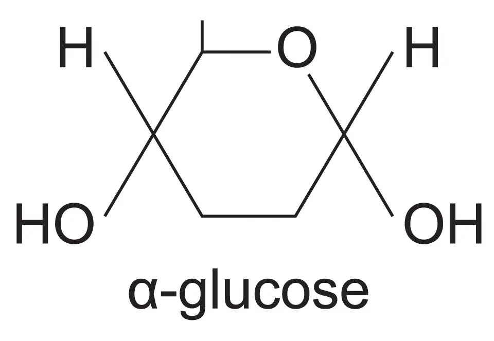
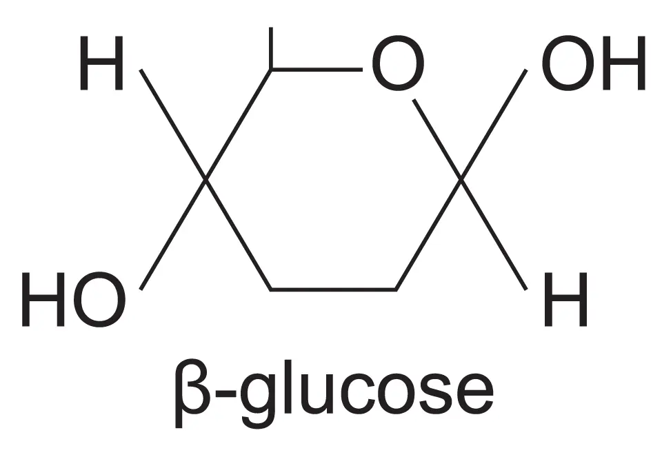
Monosaccharides: the simplest carbohydrates. Examples: glucose (C6H12O6), fructose, galactose. Both alpha (α) and beta (β) glucose have the same molecular formula but differ in the position of the -OH group on carbon 1.

α-glucose ring structure
α-glucose: -OH on C1 points down
β-glucose ring structure
β-glucose: -OH on C1 points up
α-glucose
-OH group on C1 points downward. Forms starch and glycogen via 1,4 and 1,6 glycosidic bonds. Makes coiled, branched structures.
β-glucose
-OH group on C1 points upward. Alternating units rotate 180° to form straight chains. Forms cellulose. Chains linked by hydrogen bonds into microfibrils.

Disaccharides (formed by condensation of two monosaccharides via a glycosidic bond):

Maltose
α-glucose + α-glucose. Produced by amylase digestion of starch. Reducing sugar.
Sucrose
α-glucose + fructose. Main transport sugar in plants. Non-reducing sugar.
Lactose
Glucose + galactose. Found in mammalian milk. Reducing sugar.

Polysaccharides:

PolysaccharideMonomerBondsStructureFunction
Starch (amylose)α-glucose1,4 onlyUnbranched, coiled helixEnergy storage in plants
Starch (amylopectin)α-glucose1,4 and 1,6BranchedEnergy storage in plants
Glycogenα-glucose1,4 and 1,6Highly branchedEnergy storage in animals (liver and muscle)
Celluloseβ-glucose1,4 onlyStraight chains, H-bonds between chains form microfibrilsStructural: plant cell walls

Tests: Benedict's reagent (reducing sugars: blue to brick-red precipitate on heating). Non-reducing sugars: hydrolyse with HCl first, neutralise, then Benedict's. Iodine solution: starch gives blue-black colour.

Starch and glycogen are suitable as storage molecules because they are insoluble (do not affect osmosis), compact, and can be rapidly hydrolysed to release glucose. Their branched structure provides many free ends for simultaneous enzyme action.

1.2  Lipids

Triglyceride
One glycerol + three fatty acids joined by ester bonds (condensation). Hydrophobic; used for energy storage, thermal insulation, and buoyancy.
Phospholipid
One glycerol + two fatty acids + phosphate group. Hydrophilic head (phosphate) and hydrophobic tail (fatty acids). Forms the phospholipid bilayer of membranes.
Saturated fatty acid
No C=C double bonds. Straight chain; molecules pack tightly. Higher melting point. Found in animal fats (solid at room temperature).
Unsaturated fatty acid
One or more C=C double bonds. Kinked chain; prevents tight packing. Lower melting point. Found in plant oils (liquid at room temperature).

Test for lipids: emulsion test - dissolve sample in ethanol, add to water; a cloudy white emulsion confirms lipid presence.

Per gram, lipids release more than twice the energy of carbohydrates (approximately 39 kJ g-1 vs 17 kJ g-1). They are also lighter per unit energy (no water of hydration), making them ideal for long-term energy storage.

1.3  Proteins and enzymes

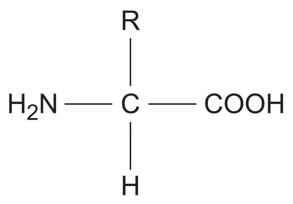
Amino acids are the monomers of proteins. Each has an amino group (-NH2), a carboxyl group (-COOH), a hydrogen atom, and a variable R group attached to a central carbon. A peptide bond forms by condensation between the amino group of one amino acid and the carboxyl group of another.

General structure of an amino acid
General amino acid structure: R group varies between the 20 common amino acids
Primary structure
The sequence of amino acids in the polypeptide chain. Determined by the gene. All higher levels of structure depend on it.
Secondary structure
α-helix or β-pleated sheet. Formed by hydrogen bonds between the -C=O and -N-H groups of the peptide backbone.
Tertiary structure
The precise 3D folding of a single polypeptide. Held by hydrogen bonds, ionic bonds, disulfide bridges, and hydrophobic interactions between R groups.
Quaternary structure
Two or more polypeptide subunits associated together. Example: haemoglobin (4 subunits) and collagen (3 polypeptide chains).

Test for proteins: biuret test - add sodium hydroxide (NaOH) solution, then a few drops of dilute copper(II) sulfate (CuSO4) solution. A purple/violet colour confirms the presence of peptide bonds. Blue = negative result (no protein).


Enzymes are biological catalysts. They lower the activation energy of reactions by forming a temporary enzyme-substrate complex at the active site.

Lock and key model (historical)
Active site has a fixed, rigid shape exactly complementary to the substrate. Substrate fits like a key in a lock. Now considered oversimplified - it cannot explain why some similarly-shaped molecules fail to act as substrates.
Induced fit model (current)
The active site changes shape slightly to mould around the substrate as it binds, forming a closer fit. The current accepted model. Explains substrate specificity more accurately and accounts for enzyme-inhibitor interactions.
Competitive inhibitor
Has a similar shape to the substrate and binds to the active site, forming an enzyme-inhibitor complex. Reversible: the inhibitor can dissociate. Substrate and inhibitor compete - increasing substrate concentration reduces the effect by outcompeting the inhibitor. Vmax is eventually reached at high enough substrate concentrations.
Non-competitive inhibitor
Binds to an allosteric site (separate from the active site). Causes a conformational change to the active site so the substrate can no longer bind. Increasing substrate concentration has no effect: the inhibitor does not compete for the active site. Vmax is reduced even at saturating substrate concentrations.

End-product inhibition (feedback inhibition): the final product of a metabolic pathway acts as a non-competitive inhibitor of an earlier enzyme in the pathway. This prevents overproduction and conserves resources. Example: amino acid synthesis pathways.

Factors affecting enzyme activity:

  • Temperature: increases rate up to optimum; above optimum the enzyme denatures - permanently changes the shape of the active site
  • pH: extreme pH denatures the enzyme; changes ionisation of R groups in the active site
  • Substrate concentration: increases rate until all active sites are occupied and all enzymes are saturated
  • Enzyme concentration: increases rate if substrate is in excess

Denaturation changes the tertiary structure permanently (breaks hydrogen bonds, ionic bonds, and disulfide bridges); this changes the shape of the active site so the substrate can no longer bind. Denaturation is not the same as the enzyme simply slowing down at low temperatures.

1.4  Nucleic acids and DNA replication

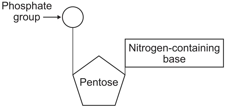
Nucleotides are the monomers of nucleic acids. Each consists of a pentose sugar, a phosphate group, and a nitrogenous base. Nucleotides join by phosphodiester bonds (condensation between the phosphate of one and the sugar of the next).

Structure of a nucleotide
A nucleotide: phosphate group + pentose sugar + nitrogen-containing base
DNA
Double helix; deoxyribose sugar; bases A, T, C, G. A pairs with T (2 H-bonds); C pairs with G (3 H-bonds). Antiparallel strands (5' to 3' and 3' to 5'). Stable; long-term information storage.
mRNA
Single stranded; ribose sugar; bases A, U, C, G (uracil replaces thymine). Carries genetic information from nucleus to ribosome. Short-lived.
tRNA
Single stranded but folded; cloverleaf shape with stem-loop structure. Anticodon at one end; amino acid attachment site at the other (CCA 3' end). Brings specific amino acids to the ribosome.
Chargaff's rules
In double-stranded DNA: %A = %T and %C = %G. A consequence of complementary base pairing. Useful for calculations: if %A = 30%, then %T = 30%, and %C = %G = 20%.

DNA replication is semi-conservative: each new DNA molecule contains one original (parental) strand and one newly synthesised strand.

  1. DNA helicase breaks the hydrogen bonds between base pairs and unwinds the double helix, creating two template strands
  2. Free DNA nucleotides are attracted to their complementary base pairs on the template strand (A with T, C with G)
  3. A primer is required to provide a free 3' end; DNA polymerase can only add nucleotides to an existing 3' end, not start a new strand from scratch
  4. DNA polymerase adds free nucleotides to the 3' end of the primer, forming phosphodiester bonds via condensation, working in the 5' to 3' direction
  5. Two identical double-stranded DNA molecules result, each with one original and one new strand

Semi-conservative replication was confirmed by the Meselson-Stahl experiment using heavy nitrogen (15N) and light nitrogen (14N). After one generation in 14N medium, all DNA had intermediate density (one strand each). After two generations, half was intermediate and half light density.

1.5  ATP, water and inorganic ions

ATP (adenosine triphosphate) consists of adenine + ribose + three phosphate groups. It is the universal energy currency of cells.

Structure of ATP
ATP: adenine (base) + ribose (pentose sugar) + three phosphate groups (P-P-P)
Hydrolysis: ATP + H2O → ADP + Pi + energy (catalysed by ATP hydrolase) Regeneration: ADP + Pi → ATP (condensation reaction; catalysed by ATP synthase)

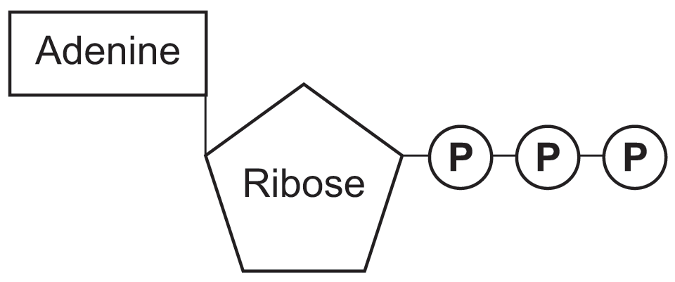
ATP is suitable as an energy currency because: it releases energy in small, manageable amounts; it is soluble and easily transported; it cannot pass out of the cell; it is immediately usable without further digestion. The inorganic phosphate (Pi) released during hydrolysis can phosphorylate other compounds, activating them for metabolic reactions.


Water's biological importance stems from its dipole nature (slightly positive H, slightly negative O), which enables hydrogen bonding between molecules:

High specific heat capacity
Large amounts of energy required to raise temperature. Buffers temperature changes in organisms and aquatic environments.
High latent heat of vaporisation
Evaporation of water requires a lot of energy, providing effective cooling (sweating, transpiration).
Solvent properties
Polar molecules and ions dissolve readily in water. Enables metabolic reactions and transport of substances in blood and cell cytoplasm.
Cohesion and surface tension
Hydrogen bonds between water molecules allow water columns to be pulled up xylem (cohesion-tension). Surface tension supports small organisms on water.
Transparent
Water allows light to pass through, enabling photosynthesis in aquatic organisms and in plant cells. Light penetrates to depth in lakes and oceans, supporting aquatic ecosystems.
Metabolite
Water is a reactant or product in many metabolic reactions. It is a product of condensation reactions (e.g. forming peptide bonds, glycosidic bonds) and a reactant in hydrolysis reactions (e.g. digestion) and photosynthesis.

Key inorganic ions:

Fe2+ (iron)
Component of haem group in haemoglobin; binds oxygen for transport in blood.
Ca2+ (calcium)
Bone and teeth structure; muscle contraction (releases from SR); enzyme cofactor; cell signalling.
Na+ (sodium)
Co-transport of glucose and amino acids across epithelium; nerve impulse transmission; osmotic balance.
PO43- (phosphate)
Forms the sugar-phosphate backbone of DNA and RNA. Component of ATP and phospholipids. Phosphorylation activates molecules in respiration and photosynthesis.
NO3- (nitrate)
Source of nitrogen for plants; used to synthesise amino acids and nucleotides.
H+ (hydrogen)
The concentration of H+ ions determines the pH of a solution. High [H+] = low pH (acidic); low [H+] = high pH (alkaline). pH changes alter the ionisation of R groups in enzyme active sites, affecting enzyme activity and tertiary structure.

Topic 2 - Cells

2.1  Cell structure: prokaryotes and eukaryotes

FeatureProkaryotic cellEukaryotic cell
Size1-10 μm10-100 μm
NucleusNo membrane-bound nucleus; naked circular DNA in nucleoid regionMembrane-bound nucleus with linear DNA and histones
OrganellesNo membrane-bound organellesMembrane-bound organelles (mitochondria, ER, Golgi etc.)
Ribosomes70S (smaller)80S (larger); 70S in mitochondria and chloroplasts
Cell wallMurein (peptidoglycan)Cellulose (plants), chitin (fungi), absent in animals
DNACircular, naked (no histones); may have plasmidsLinear, associated with histone proteins

Key eukaryotic organelles:

Nucleus
Double membrane (nuclear envelope) with nuclear pores. Nucleolus (synthesises rRNA and ribosomes). Contains chromosomes (DNA + histones). Controls cell activity.
Mitochondria
Double membrane; inner membrane folded into cristae (increases SA). Matrix contains enzymes, circular DNA, and 70S ribosomes. Site of aerobic respiration (Krebs cycle and oxidative phosphorylation).
Rough ER
Studded with ribosomes on its outer surface. Folds and processes proteins synthesised by ribosomes. Transports proteins in vesicles to the Golgi apparatus.
Golgi apparatus
Stack of flattened membranes. Modifies, packages, and sorts proteins (e.g. adds carbohydrate chains to make glycoproteins). Produces secretory vesicles and lysosomes.
Lysosomes
Small vesicles containing hydrolytic enzymes. Digest engulfed bacteria (phagocytosis), worn-out organelles (autophagy), and foreign material.
Chloroplast
Double membrane; thylakoids stacked into grana; fluid-filled stroma. Contains circular DNA and 70S ribosomes. Site of photosynthesis.

Microscopy:

Light microscope
Max magnification ~1500x; resolution ~200 nm. Uses visible light. Specimens may be stained. Can view living cells. Used for tissues and organelles down to ~1 μm.
Electron microscope
Max magnification ~500,000x; resolution ~0.1 nm. Uses electrons. Specimens must be dead and in a vacuum. TEM: thin sections, detailed internal structure. SEM: surface, 3D image.
Magnification = image size / actual size Resolution = minimum distance between two points that can be distinguished as separate

Magnification and resolution are different. A blurry photo can be magnified further but the detail does not improve (resolution is limited). Electron microscopes have far greater resolution than light microscopes, allowing sub-cellular structures to be seen clearly.


Viruses are acellular and non-living. They cannot carry out metabolism, grow, or reproduce independently. Structure of a virus particle (virion):

Genetic material
Either DNA or RNA (never both). May be single- or double-stranded. Carries instructions for making new virus particles using the host cell's machinery.
Capsid
Protein coat surrounding and protecting the genetic material. Made of protein subunits called capsomeres. Shape varies: helical, icosahedral, or complex.
Attachment proteins
Glycoproteins on the outer surface. Complementary to specific receptor proteins on the host cell surface. Determine host cell specificity (which cells the virus can infect).

Cell fractionation and ultracentrifugation isolate specific organelles from cells for biochemical study:

  1. Tissue is homogenised in ice-cold isotonic buffer. Ice-cold: reduces enzyme activity; isotonic: prevents osmotic damage to organelles; buffer: maintains stable pH
  2. Homogenate is filtered to remove intact cells and large debris
  3. Filtered solution is centrifuged at progressively increasing speeds. Denser and larger organelles pellet first:
~1,000 g - nuclei pellet first (largest/densest) ~10,000 g - mitochondria and chloroplasts pellet ~100,000 g - ribosomes and ER membrane fragments pellet

All three conditions (cold, isotonic, buffered) are needed simultaneously. Cell fractionation allowed organelles to be isolated and their biochemical roles confirmed directly - e.g. isolated mitochondria confirmed that aerobic respiration occurs in mitochondria.

2.2  The cell cycle, mitosis and meiosis

The cell cycle has two main phases:

G1 (Gap 1)
Cell grows; proteins synthesised; organelles replicated. Preparation for DNA replication.
S phase (Synthesis)
DNA replication occurs. Each chromosome consists of two identical chromatids joined at the centromere.
G2 (Gap 2)
Further cell growth; organelles replicate; preparation for mitosis.

Mitosis produces two genetically identical daughter cells (same chromosome number as parent). Used for growth, repair, and asexual reproduction.

Prophase
Chromosomes condense and become visible. Spindle fibres form from centrioles. Nuclear envelope breaks down.
Metaphase
Chromosomes align at the equator (metaphase plate). Spindle fibres attach to centromeres.
Anaphase
Centromeres split. Sister chromatids pulled to opposite poles by shortening spindle fibres.
Telophase + Cytokinesis
Nuclear envelopes reform around each set of chromosomes. Cell divides into two identical daughter cells.

Meiosis produces four genetically non-identical haploid cells. Involves two successive divisions. Used for gamete production. Sources of genetic variation:

  • Crossing over: homologous chromosomes exchange segments at chiasmata during prophase I. Creates new combinations of alleles
  • Independent assortment: homologous pairs line up randomly at the equator during metaphase I; each combination of chromosomes is possible

Mitosis and meiosis are commonly confused. Key distinctions: mitosis = 2 diploid daughter cells, genetically identical; meiosis = 4 haploid daughter cells, genetically unique. Only meiosis involves crossing over and independent assortment.


Binary fission in prokaryotes (not mitosis - no spindle, no linear chromosomes):

  1. The circular DNA replicates; any plasmids also replicate
  2. The cell elongates; the two DNA copies are pulled to opposite ends of the cell
  3. The cytoplasm divides, producing two daughter cells - each with one copy of the circular DNA and a variable number of plasmid copies

Virus replication: viruses do not divide - they are non-living. After attachment proteins bind to the host cell surface, viral nucleic acid is injected into the host cell. The host cell's own ribosomes and enzymes replicate the viral nucleic acid and synthesise viral proteins. New virions are assembled and released by lysis or budding.

Cancer: mitosis is normally tightly regulated by genes. Mutations in these control genes can cause uncontrolled cell division, producing a tumour. If tumour cells spread to other tissues (metastasis), this is cancer. Many cancer treatments target rapidly dividing cells - e.g. chemotherapy disrupts the cell cycle; radiotherapy damages DNA in tumour cells.

Mitotic index = (number of cells in mitosis) / (total number of cells observed) A high mitotic index indicates rapid cell division (e.g. growing tissue, tumour cells).

2.3  Transport across cell membranes

The fluid mosaic model describes the cell surface membrane: a phospholipid bilayer (hydrophilic heads face outward, hydrophobic tails face inward) with proteins embedded throughout. Cholesterol stabilises fluidity. Glycoproteins and glycolipids act as cell surface receptors and antigens.


Simple diffusion
Passive; down a concentration gradient; through the lipid bilayer. Only for small, non-polar, lipid-soluble molecules (e.g. O2, CO2, ethanol). Rate proportional to concentration gradient.
Facilitated diffusion
Passive; down a concentration gradient; via channel proteins (for ions) or carrier proteins (for larger polar molecules). Specific to particular molecules. Can be saturated at high concentrations.
Osmosis
The movement of water molecules from a region of higher water potential to a region of lower water potential through a partially permeable membrane. Water potential (Ψ) = Ψs + Ψp. Pure water has Ψ = 0; adding solute makes Ψ more negative.
Active transport
Against the concentration gradient; requires ATP and carrier proteins. Example: Na+/K+ ATPase pump. Co-transport: uses Na+ gradient (established by active transport) to drive uptake of glucose and amino acids.
Endocytosis
Cell membrane engulfs large particles or droplets to form a vesicle. Phagocytosis (solid particles); pinocytosis (liquids). Requires ATP.
Exocytosis
Vesicles from Golgi fuse with the cell surface membrane and release their contents outside the cell. Used for secretion of proteins (e.g. hormones, digestive enzymes).

Water potential must be understood carefully: it is always negative or zero. A cell placed in a hypertonic solution (lower water potential than cell) will lose water by osmosis and shrink (crenation in animal cells; plasmolysis in plant cells). A cell in a hypotonic solution gains water (lysis in animal cells; turgid but protected by cell wall in plants).

2.4  Cell recognition and the immune system

Each cell type carries specific molecules on its surface - primarily proteins and glycoproteins - that identify it. The immune system uses these to distinguish self from non-self, identifying: pathogens, cells from other organisms of the same species, abnormal body cells, and toxins.

Antigen (definition)
A molecule (usually a protein or glycoprotein) on the surface of a cell or pathogen that is recognised as foreign by the immune system and triggers an immune response. Self antigens on body cells are recognised and not attacked.
Antigen variability
Some pathogens (e.g. influenza) frequently change their surface antigens (antigenic variation). Existing memory cells and antibodies no longer recognise the new antigen. This is why flu vaccines must be updated annually and why some diseases are difficult to eliminate.

Phagocytosis:

  1. Phagocyte is attracted to pathogen by chemical signals (chemotaxis)
  2. Phagocyte engulfs the pathogen by endocytosis, enclosing it in a phagosome
  3. Lysosomes fuse with the phagosome and release hydrolytic enzymes (lysozymes) that digest the pathogen
  4. Antigens from the digested pathogen are displayed on the phagocyte's cell surface - it becomes an antigen-presenting cell (APC)

The cellular response (T lymphocytes):

Helper T cells (TH)
Activated when their surface receptors bind to antigens displayed by APCs. Release cytokines that stimulate: cytotoxic T cells (to kill infected cells), B cells (to produce antibodies), and phagocytes (to increase phagocytosis). TH cells coordinate both arms of the immune response.
Cytotoxic T cells (TC)
Kill infected body cells, cancer cells, and transplanted cells by inducing apoptosis (programmed cell death). They recognise foreign antigens displayed on the surface of host cells. Activated by cytokines from helper T cells.

The humoral response (B lymphocytes):

Clonal selection and expansion
The B cell whose surface antibodies are complementary to the antigen binds to it. Stimulated by helper T cell cytokines, this B cell undergoes rapid mitosis (clonal expansion), producing many identical B cells that differentiate into plasma cells and memory cells.
Antibody (definition and structure)
A protein (immunoglobulin) produced by plasma cells with a specific antigen-binding site complementary to one antigen. Y-shaped: two heavy + two light polypeptide chains linked by disulfide bridges. Variable regions form the two antigen-binding sites; constant region determines class and effector function.
Plasma cells
Differentiated B cells that produce and secrete large quantities of specific antibodies into the blood and lymph. Short-lived (days to weeks). Generate the primary immune response antibody titre.
Memory cells
Long-lived B and T lymphocytes persisting after infection. Carry the specific antigen receptor. On re-exposure to the same antigen, they divide rapidly into large numbers of plasma/effector cells - producing a faster, stronger secondary immune response, usually clearing infection before symptoms develop.

Antigen-antibody complex and pathogen destruction:

Agglutination
Each antibody has two antigen-binding sites. Antibodies cross-link multiple pathogens into large clumps. Clumped pathogens cannot infect cells and are more easily engulfed by phagocytes.
Opsonisation
Antibodies coat the surface of pathogens. Phagocytes have receptors for the antibody constant region and preferentially engulf opsonised pathogens - greatly increasing the rate of phagocytosis.

Primary immune response
First exposure to an antigen. Slow to develop (days to weeks) as clonal selection and expansion take time. Antibody titre rises slowly to a relatively low level. Symptoms may develop. Memory cells are formed.
Secondary immune response
Re-exposure to the same antigen. Memory cells divide rapidly into large numbers of plasma cells. Antibody titre rises much faster and to a much higher level. Pathogen usually eliminated before symptoms appear.

Active immunity
The immune system produces its own antibodies after exposure to an antigen. Produces memory cells - long-lasting protection. Natural active: through infection. Artificial active: through vaccination. Takes time to develop initially.
Passive immunity
Antibodies are received from an external source. No memory cells are produced - protection is temporary (weeks) as the antibodies are broken down. Natural passive: maternal antibodies via placenta and breast milk. Artificial passive: injection of specific antibodies (e.g. anti-tetanus immunoglobulin).

Vaccines and herd immunity: a vaccine contains an antigen (killed/attenuated pathogen, protein subunit, or mRNA) that stimulates a primary immune response and memory cell formation without causing disease. Herd immunity: when a sufficiently high proportion of a population is immune, chains of infection break down and the pathogen cannot spread - protecting unvaccinated individuals (e.g. newborns, immunocompromised) who cannot receive vaccines.


HIV and AIDS:

HIV structure
Retrovirus. Genetic material: RNA (two copies). Contains reverse transcriptase enzyme. Surrounded by a protein capsid, then a lipid envelope studded with glycoprotein attachment proteins (gp120) that are complementary to CD4 receptors on helper T cells.
HIV replication and AIDS
HIV attachment proteins bind to CD4 receptors on helper T cells. RNA is reverse-transcribed to DNA by reverse transcriptase, then inserted into the host genome. Helper T cell produces viral RNA and proteins; new virions bud off. Progressive destruction of TH cells eventually causes AIDS - the immune system fails and the patient becomes vulnerable to opportunistic infections.

Why antibiotics are ineffective against viruses: antibiotics target bacterial structures - cell wall synthesis, 70S ribosomes, metabolic enzymes. Viruses have no cell wall, no ribosomes of their own, and no independent metabolism; they use the host cell's machinery. There is no bacterial target for antibiotics to act on.


Monoclonal antibodies are identical antibodies produced by a single clone (hybridoma cell = B lymphocyte fused with a myeloma cancer cell, combining antibody production with immortality). Uses:

Targeting medication
A therapeutic drug or radioactive molecule is attached to a monoclonal antibody specific to an antigen on target cells (e.g. cancer cells). The antibody delivers the drug directly to the target, minimising damage to healthy tissue. Example: Herceptin targets HER2 receptors on breast cancer cells.
Medical diagnosis
Monoclonal antibodies detect specific antigens in patient samples. Examples: pregnancy tests (detect hCG hormone); blood typing; detection of pathogens or tumour markers; HIV testing. Basis of the ELISA test.

ELISA test (enzyme-linked immunosorbent assay): detects the presence or quantity of a specific antigen. Antigen in sample binds to surface - primary antibody (complementary to antigen) is added - secondary antibody (with enzyme attached) binds to primary antibody - substrate is added - the enzyme converts substrate to a coloured product. Colour development confirms presence of antigen; intensity indicates quantity.

Ethical issues: religious/cultural objections to vaccination; rare adverse reactions balanced against population benefit; debate over compulsory vaccination; herd immunity obligations vs individual choice; use of animals in producing monoclonal antibodies; high cost limiting access in lower-income countries; uncertainty about long-term effects of newer technologies (e.g. mRNA vaccines).

Active vs passive: the key difference is memory cells. Active immunity creates them (long-lasting); passive immunity does not (temporary). HIV destroys helper T cells - these coordinate both the cellular and humoral responses, so losing them progressively cripples the entire immune system, leading to AIDS.

Topic 3 - Organisms exchange substances with their environment

3.1 – 3.2  Exchange surfaces and gas exchange

As organisms increase in size, their surface area to volume (SA:V) ratio decreases. Large organisms cannot exchange substances fast enough by diffusion alone and require specialised exchange surfaces with the following features:

  • Large surface area to maximise rate of diffusion
  • Thin walls (short diffusion distance)
  • Moist surface (gases dissolve before diffusing)
  • Good blood supply (maintains concentration gradient)
  • Ventilation (maintains concentration gradient on air side)

Gas exchange in mammals (alveoli): millions of alveoli provide an enormous total surface area (~70 m2). Walls are one cell thick; surrounded by capillary network. Surfactant reduces surface tension to prevent collapse.

Ventilation: Inhalation - external intercostal muscles and diaphragm contract; thorax volume increases; pressure falls below atmospheric; air moves in. Exhalation - muscles relax; volume decreases; pressure rises; air moves out. (Forced exhalation: internal intercostal muscles contract.)


Single-celled organisms
Amoeba and other single-celled organisms exchange gases directly across the cell surface membrane. Their large SA:V ratio (due to small size) means diffusion alone is fast enough. No specialised gas exchange surface is needed.
Insect gas exchange
Tracheal system: spiracles (controlled openings) lead to tracheae, branching into tracheoles that reach every cell. Gas exchange by diffusion. Spiracles close to reduce water loss - a structural compromise: open spiracles = better gas exchange but greater water loss.
Fish gas exchange
Water flows over gill lamellae in the opposite direction to blood flow (counter-current exchange). Maintains a diffusion gradient along the entire gill, allowing up to 80% O2 extraction from water (parallel flow would only achieve ~50%).
Dicotyledonous plant gas exchange
CO2 enters through stomata in the leaf epidermis, surrounded by guard cells. Large air spaces in the spongy mesophyll maximise surface area. Guard cells open stomata in light (K+ influx increases solute concentration - water enters by osmosis - cell becomes turgid).
Xerophytic plant adaptations
Xerophytes (plants adapted to dry conditions) reduce water loss at the cost of reduced gas exchange: thick waxy cuticle; sunken or rolled stomata (trap humid air, reduce water potential gradient); fewer stomata; hairy leaves (trap moist air layer). Structural compromise: the same adaptations that reduce water loss also reduce CO2 entry rate.

Pulmonary ventilation rate (PVR):

PVR = tidal volume × breathing rate PVR: volume of air moved in/out of lungs per minute (dm³ min¹) Tidal volume: volume of air per breath (dm³) Breathing rate: number of breaths per minute

The counter-current system in fish gills maintains a diffusion gradient along the entire gill surface - blood is always less saturated than the water next to it. A parallel flow system would reach equilibrium halfway, reducing extraction to ~50%. Xerophyte adaptations always involve a compromise: reducing water loss also restricts gas exchange efficiency.

3.3  Digestion and absorption

EnzymeTypeSubstrateProduct(s)Location
Salivary amylaseCarbohydraseStarchMaltose (and dextrins)Mouth
Pancreatic amylaseCarbohydraseStarchMaltose (and dextrins)Small intestine
MaltaseDisaccharidaseMaltoseGlucoseSmall intestine (brush border)
PepsinEndopeptidase (cleaves internal peptide bonds)ProteinsPolypeptidesStomach (pH 2)
TrypsinEndopeptidase (cleaves internal peptide bonds)Proteins/polypeptidesSmaller peptidesSmall intestine
ExopeptidasesExopeptidase (cleaves terminal amino acids)PolypeptidesShorter peptides/amino acidsSmall intestine
DipeptidasesMembrane-bound exopeptidaseDipeptidesAmino acidsSmall intestine (brush border)
LipaseLipaseTriglyceridesFatty acids + monoglyceridesSmall intestine

Bile (produced by liver, stored in gall bladder) emulsifies fats into smaller droplets, increasing surface area for lipase action. Fatty acids and monoglycerides are incorporated into micelles (with bile salts), which carry them to the epithelial surface where they are released and absorbed.

Absorption in the ileum: villi (and microvilli/brush border on epithelial cells) greatly increase surface area. Glucose and amino acids absorbed by co-transport with Na+ (Na+ enters with glucose via a co-transporter; Na+ pumped back out by Na+/K+ ATPase). Fatty acids and monoglycerides diffuse out of micelles into epithelial cells, are reassembled into triglycerides, packaged into chylomicrons, and enter the lacteals (lymphatic system).

Co-transport relies on the sodium gradient established by active transport of Na+ out of the cell. Glucose therefore enters against its own concentration gradient indirectly, using energy spent maintaining the Na+ gradient.

3.4  Mass transport in animals

Arteries
Carry blood away from the heart. Thick, elastic, muscular walls. High pressure. No valves. Small lumen relative to wall thickness.
Veins
Return blood to the heart. Thin walls; large lumen. Low pressure. Valves prevent backflow. Skeletal muscle contraction assists flow.
Capillaries
Site of exchange. Walls one cell thick (endothelium only). Very small lumen. Substances exchange by diffusion, osmosis, and filtration.

Tissue fluid formation: at the arterial end of a capillary, hydrostatic pressure (blood pressure) is high, forcing fluid out. Plasma proteins lower the water potential of the blood, so at the venous end (where hydrostatic pressure has fallen), water moves back into the capillary by osmosis down a water potential gradient. Excess fluid drains into the lymphatic system.


The heart: right side pumps deoxygenated blood to lungs (pulmonary circulation); left side pumps oxygenated blood to body (systemic circulation). The left ventricle has thicker walls (generates higher pressure for systemic circulation).

Cardiac cycle: atrial systole (atria contract, blood enters ventricles) → ventricular systole (ventricles contract, blood ejected) → diastole (all chambers relax, heart refills). Atrioventricular valves prevent backflow into atria; semilunar valves prevent backflow from arteries.

Cardiac output = heart rate × stroke volume (unit: cm3 min-1 or L min-1)

Haemoglobin is a quaternary protein with four subunits, each containing a haem group with an Fe2+ ion that binds one O2 molecule (so 4 O2 per haemoglobin). The oxygen dissociation curve is S-shaped (sigmoid) due to cooperative binding (binding of one O2 makes subsequent binding easier).

Bohr effect
Increased CO2 (lowers pH) shifts the dissociation curve to the right: haemoglobin has a lower affinity for O2 and releases it more readily to respiring tissues.
Fetal haemoglobin
Higher affinity for O2 than adult haemoglobin (curve shifted left). Allows fetal blood to load O2 from maternal blood across the placenta at relatively low pO2.

The sigmoid shape of the oxygen dissociation curve is biologically significant: haemoglobin loads O2 efficiently in the lungs (high pO2, steep part of curve) and unloads efficiently in the tissues (low pO2). The cooperative binding mechanism makes it far more effective than a simple linear relationship would be.

3.5  Mass transport in plants

Xylem
Carries water and mineral ions from roots to leaves. Dead cells with lignified walls; no cytoplasm. Vessel elements and tracheids. Water moves by cohesion-tension mechanism.
Phloem
Carries organic solutes (mainly sucrose) from source to sink. Living sieve tube elements with companion cells. Moves by the mass flow hypothesis.

Transpiration and cohesion-tension theory:

  1. Water evaporates from mesophyll cells into air spaces and exits through stomata (transpiration)
  2. Water potential in leaf mesophyll falls; water moves from xylem by osmosis
  3. Cohesion between water molecules (hydrogen bonds) creates a continuous water column under tension
  4. Tension is transmitted down the xylem, pulling water up from roots
  5. Water enters root hair cells by osmosis (lower water potential in root cells than soil solution)

Factors affecting transpiration rate: temperature (increases evaporation); humidity (reduces gradient if high); wind speed (removes water vapour, steepens gradient); light (stomata open); leaf area.


Translocation (mass flow hypothesis): sugars are actively loaded into phloem at the source (e.g. leaf) raising the solute concentration. Water enters phloem by osmosis, increasing hydrostatic pressure. This drives mass flow of solutes towards the sink (e.g. roots, growing regions) where sugars are unloaded.


Evidence for and against the mass flow hypothesis:

Ringing experiments
A ring of bark (including phloem) is removed from a stem. Sugars accumulate above the ring and cannot pass below it - confirming that phloem (not xylem) transports organic solutes. The xylem continues to function (water still moves upward).
Tracer experiments
Radioactively labelled CO2 (14C) is supplied to a leaf in light. The 14C is fixed into sugars and can be detected moving through the phloem away from the leaf - in both directions (toward growing tips and roots), consistent with mass flow from source to sink.

Evidence against mass flow: all solutes in phloem sap move at the same speed (consistent with mass flow), but different sugars and amino acids are sometimes found at different concentrations at different points - suggesting selective loading/unloading rather than simple bulk flow. The mechanism of unloading at sinks is also more complex than simple mass flow predicts.

Water movement in xylem is passive (no ATP); translocation in phloem requires ATP (active loading at source). Evidence for mass flow: companion cells have many mitochondria (active loading); metabolic inhibitors stop translocation; phloem sap is under positive pressure (aphid stylet experiments show sap exudes). Xylem carries water up; phloem carries organic solutes in both directions.

Topic 4 - Genetic information, variation and relationships between organisms

4.1  DNA, genes and chromosomes

Gene
A sequence of DNA bases on a chromosome that codes for the amino acid sequence of a polypeptide (or a functional RNA molecule).
Allele
One of two or more alternative forms of a gene, occupying the same locus on homologous chromosomes.
Locus
The specific position of a gene on a chromosome. The locus is the same in all individuals of a species; the allele at that locus may differ.
Homologous chromosomes
Matching pairs of chromosomes with the same genes at the same loci (but potentially different alleles). One from each parent. Same length and centromere position.

DNA in eukaryotes is associated with histone proteins. DNA wraps around histones to form nucleosomes, which coil further to form chromatin. The coiling allows the DNA of a human cell (~2 m of DNA) to fit inside a nucleus of ~6 μm diameter.

Organelle DNA
Mitochondria and chloroplasts each contain their own DNA. Like prokaryotic DNA, it is short, circular, and not associated with histone proteins. This is evidence for the endosymbiotic theory - that these organelles were once free-living prokaryotes.
Genome and proteome
Genome: the complete set of genes (all the DNA) in a cell. Proteome: the full range of proteins that a cell is able to produce. The proteome is larger than the genome because one gene can produce multiple proteins (through alternative splicing of exons).

Eukaryotic nuclear DNA is long, linear, and histone-associated. Organelle DNA (mitochondria, chloroplasts) is short, circular, and not histone-associated - identical in character to prokaryotic DNA. In prokaryotes, DNA is short, circular, and not histone-associated. Non-coding DNA includes introns, repetitive sequences between genes (satellite DNA), and regulatory regions.

4.2  Protein synthesis: transcription and translation

The genetic code is: triplet (3 bases = 1 codon = 1 amino acid); degenerate (multiple codons for most amino acids); non-overlapping; universal (same in almost all organisms).


Transcription (DNA → mRNA):

  1. RNA polymerase binds to the promoter region of the gene on the DNA
  2. The double helix unwinds; the template (antisense) strand is exposed
  3. Free RNA nucleotides pair with the template DNA strand by complementary base pairing (template A → mRNA U; template T → mRNA A; template C → mRNA G; template G → mRNA C); RNA polymerase joins them by phosphodiester bonds
  4. A primary (pre-mRNA) transcript is produced
  5. Splicing: introns (non-coding regions) are removed; exons (coding regions) are joined together to form mature mRNA
  6. mRNA leaves the nucleus via nuclear pores

Translation (mRNA → polypeptide):

  1. mRNA binds to a ribosome at the start codon (AUG, codes for methionine)
  2. tRNA with complementary anticodon brings the corresponding amino acid to the ribosome
  3. Peptide bonds form between adjacent amino acids (condensation); tRNA is released
  4. The ribosome moves along the mRNA (5' to 3'); continues until a stop codon (UAA, UAG, or UGA) is reached
  5. The polypeptide is released and folds into its tertiary structure

Types of gene mutation: substitution (one base replaced; may be silent, missense, or nonsense); deletion/insertion (frameshift - alters all codons downstream; usually more severe than substitution).

A silent mutation does not change the amino acid sequence (due to degeneracy of the genetic code). A nonsense mutation introduces a stop codon, producing a truncated, usually non-functional polypeptide. A missense mutation changes one amino acid, which may or may not affect protein function depending on its location and chemical properties.

4.3  Genetic diversity, selection and evolution

Genetic diversity is the range of alleles present in a population. It is the raw material for natural selection.

Sources of genetic variation
Mutations (the only source of new alleles); sexual reproduction (crossing over and independent assortment produce new allele combinations in offspring).
Natural selection
Variation exists; some variants are better adapted to the environment; better-adapted individuals survive and reproduce; pass on alleles to offspring; allele frequencies change over generations.
Genetic bottleneck
A drastic reduction in population size (e.g. disaster, hunting). Survivors may not represent the original gene pool; allele frequencies in the new population differ. Genetic diversity is reduced.
Founder effect
A small group establishes a new isolated population. The founders carry only a sample of the original gene pool; the new population has low genetic diversity. A type of genetic bottleneck.
Directional selection
One extreme phenotype is favoured. The mean of the trait shifts in one direction over generations. Example: antibiotic resistance in bacteria.
Stabilising selection
Intermediate phenotypes are favoured; extremes are selected against. Reduces variation. Example: human birth weight (very low or very high weight increases mortality).

Genetic diversity is reduced by inbreeding (increases homozygosity), genetic bottlenecks, and the founder effect. It is maintained by mutation, sexual reproduction, and migration (gene flow). Low genetic diversity makes a population more vulnerable to new diseases or environmental changes.

4.4  Species, taxonomy and biodiversity

The biological species concept: two organisms belong to the same species if they can interbreed to produce fertile offspring. Organisms are reproductively isolated from other groups.

Courtship behaviour
A necessary precursor to successful mating. Functions in species recognition: signals (visual, auditory, chemical, behavioural) are species-specific, ensuring individuals mate only with the same species and not waste gametes. Also allows assessment of mate quality (health, fitness). Examples: bird song, firefly bioluminescence patterns, pheromone signals.

Taxonomy is the classification of organisms into a hierarchy of groups based on shared characteristics:

Domain → Kingdom → Phylum → Class → Order → Family → Genus → Species (Mnemonic: Dear King Philip Came Over For Good Soup)

Binomial nomenclature: each species has a two-part Latin name: Genus (capitalised) + species (lower case). Written in italics or underlined.


Phylogenetics classifies organisms based on evolutionary relationships rather than shared physical characteristics. DNA base sequence comparisons, mRNA base sequences, and amino acid sequences in proteins provide molecular evidence for evolutionary relationships. More similarities = more recent common ancestor.


Biodiversity includes species, genetic, and ecosystem diversity. Measuring biodiversity:

Index of diversity: d = N(N-1) / Σn(n-1) where N = total number of organisms in the community n = total number of organisms of each species A higher value of d indicates greater biodiversity.

Species richness (number of species) alone ignores evenness (relative abundance). An area with 10 species of equal abundance is more diverse than one dominated by a single species.


Farming and biodiversity: intensive farming practices reduce biodiversity through: monocultures (single crop species eliminates habitat diversity); use of herbicides and pesticides (kill non-crop species and insects); removal of hedgerows (destroys habitat and wildlife corridors); drainage of wetlands; use of inorganic fertilisers (causes eutrophication). The balance between productive farming and conservation is an ongoing tension - nature reserves, hedgerow protection, and wildlife corridors are conservation measures.


Investigating genetic diversity can be done by comparing:

  • The frequency of measurable or observable characteristics within/between species
  • The base sequence of DNA
  • The base sequence of mRNA
  • The amino acid sequence of proteins encoded by DNA/mRNA

Gene technology has shifted investigations from inferring DNA differences from observable characteristics to direct sequencing of DNA - more accurate and can reveal relationships not apparent from morphology. Quantitative investigations of variation involve: collecting data from random samples; calculating the mean and standard deviation of measurements. A larger standard deviation indicates greater variation within the sample.

DNA evidence has revised many traditional taxonomic classifications. Organisms that look similar may be only distantly related (convergent evolution) while organisms with different appearances may be closely related. Molecular evidence - DNA base sequences, mRNA sequences, amino acid sequences - is now considered more reliable than morphology alone for determining evolutionary relationships.